 English

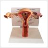
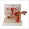

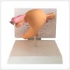
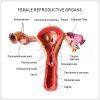
Anatomical Female Reproductive System Pathology Model for Medical Teaching
$16.50
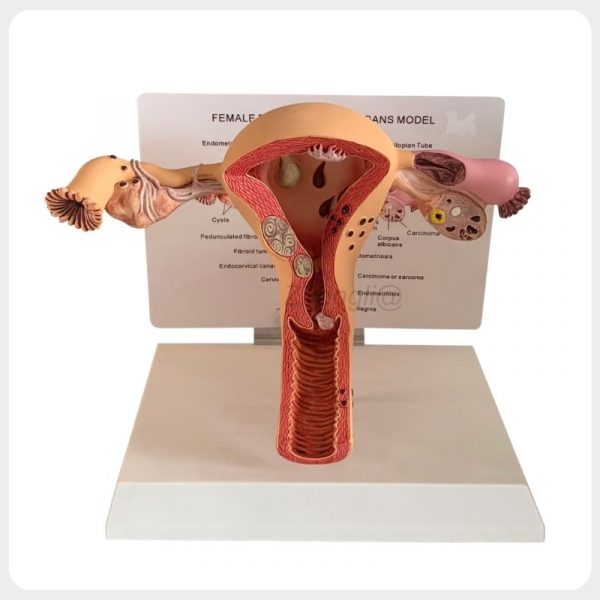
This female reproductive pathology model combines detailed uterus, ovary, fallopian tube, cervix and vaginal anatomy with multiple color-coded lesions. It provides a clear visual tool for gynecology, pathology and patient education.
1000 in stock
Product Description:
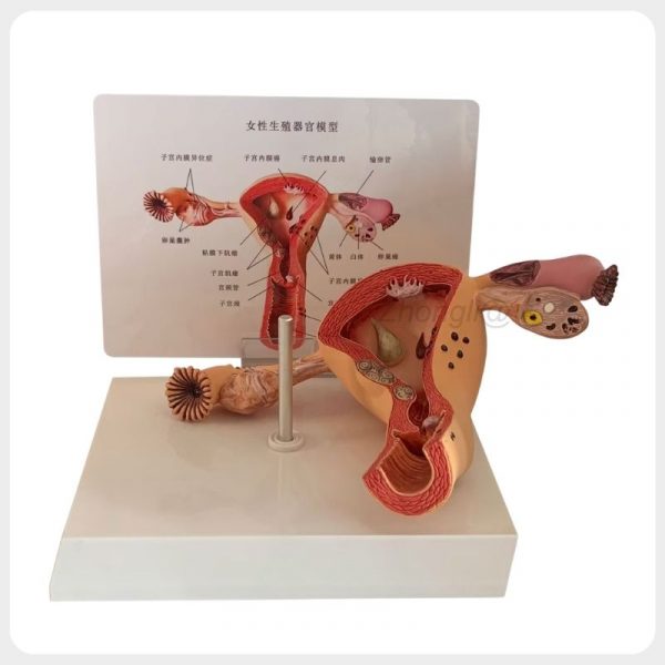
The Anatomical Female Reproductive System Pathology Model presents normal reproductive anatomy and representative pathological changes in one detailed three-dimensional display. Its cutaway uterus, bilateral ovaries, fallopian tubes, cervix and vagina help learners understand the locations and structural relationships of common gynecological diseases.
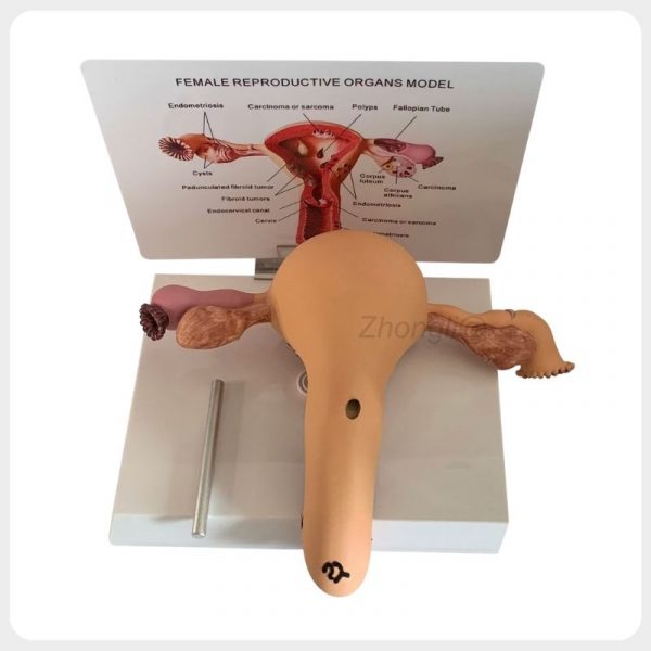
Mounted on a stable base with a labeled reference card, the model is designed for medical schools, hospitals, training institutions and professional patient education.
Key Features:
– Complete Reproductive Anatomy: Integrates the uterus, cervix, vagina, bilateral ovaries and fallopian tubes for systematic teaching.
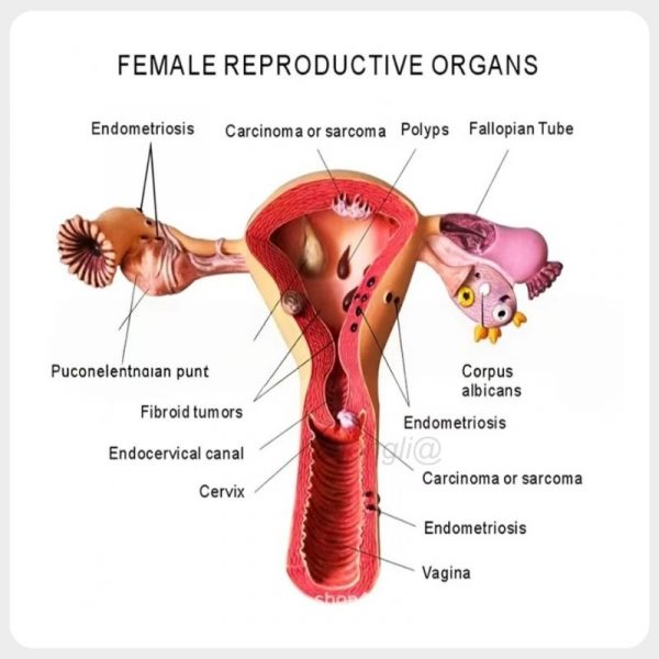
– Multiple Pathological Lesions: Demonstrates cysts, endometriosis, uterine fibroids, a pedunculated fibroid, polyps and representative malignant lesions.
– Normal and Pathological Comparison: Displays the corpus luteum and corpus albicans alongside cystic and malignant ovarian changes.
– Three-Dimensional Cutaway Design: Reveals the uterine cavity, uterine wall, cervical canal and vaginal canal more clearly than flat diagrams.
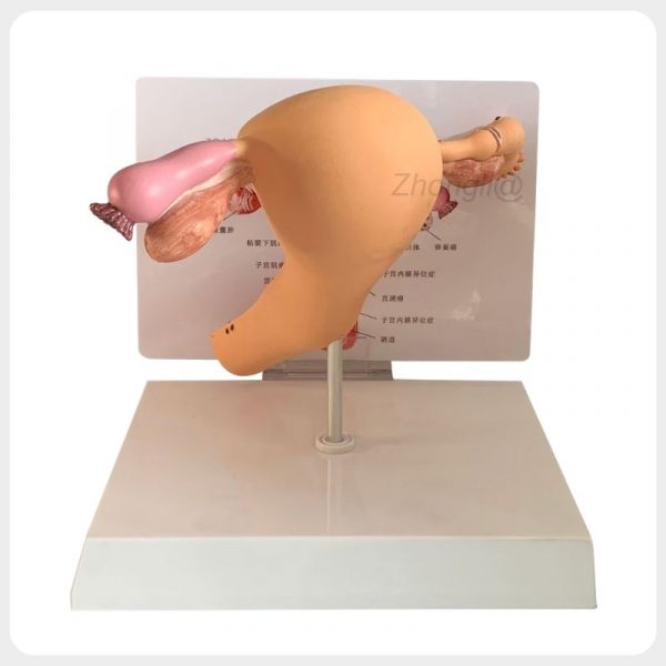
– Stable Labeled Display: The base, metal support rod and reference card support convenient classroom demonstrations and patient explanations.
Technical Specifications:
| Specification | Details |
|---|---|
| Product Name | Female Reproductive System Pathology Model |
| Model | SK-UR-002 |
| Material | Eco-Friendly PVC |
| Product Size | 18 x 15 x 20 cm |
| Product Weight | 0.4 kg |
| Scale | Anatomical Teaching Representation |
| Product Type | Pathological Anatomical Model |
| Section Design | Three-Dimensional Cutaway |
| Anatomical Structures | Uterus, Cervix, Vagina, Ovaries and Fallopian Tubes |
| Displayed Pathologies | Cysts, Endometriosis, Fibroids, Polyps and Malignant Lesions |
| Normal Ovary Structures | Corpus Luteum and Corpus Albicans |
| Display System | Base with Metal Support Rod |
| Reference System | Labeled Anatomical Card |
| Application | Gynecology, Pathology and Patient Education |
| Brand | Zhongli |
| Packaging | Individual Protective Packaging |
| Supplier Type | Manufacturer and B2B Wholesale Supplier |
Applications:
- – Female reproductive anatomy and gynecology classes in medical universities and teaching laboratories.
- – Pathology and nursing education in hospitals, nursing schools and professional training institutions.
- – Patient education in gynecology clinics, reproductive health centers and women’s health facilities.
- – Medical demonstrations, health exhibitions and reproductive disease awareness programs.
Packaging & Shipping

Packaging: The product is packed in a strong corrugated carton box to ensure safe transportation.
Inside the carton, the product is protected with foam or protective materials to prevent damage during shipping.
Standard export carton packaging is used to ensure the product arrives safely.

Global Shipping Options:
We support international express, air shipping, and sea shipping to ensure fast and reliable delivery worldwide.
Different shipping options are available depending on your order size and delivery time requirements
OEM & Customization
We support OEM and customized production based on customer requirements, including logo printing, color modification, packaging customization, and product structure adjustment.
Feel free to contact us for more details about private labeling and bulk order cooperation.
Get Our Latest Catalog
We also offer a full range of models in different sizes and structures. If you are interested, please feel free to contact us for our complete product catalog.
FAQ
Q1: Which anatomical structures are displayed on the model?
A1: The model displays the uterus, uterine cavity, uterine wall, cervix, endocervical canal, vagina, bilateral ovaries and fallopian tubes.
Q2: Which pathological conditions are represented?
A2: The model demonstrates ovarian cysts, endometriosis, uterine fibroids, a pedunculated fibroid, polyps, ovarian carcinoma and representative carcinoma or sarcoma lesions.
Q3: Does the model display normal ovarian structures?
A3: Yes. The corpus luteum and corpus albicans are displayed alongside pathological ovarian changes for comparative teaching.
Q4: Is this model suitable for patient education?
A4: Yes. Its three-dimensional and color-coded structure helps patients understand the locations of common female reproductive diseases.
Q5: What material is the model made from?
A5: The model is made from eco-friendly PVC for durable and repeated educational use.
Q6: What are the product dimensions and weight?
A6: The model measures 18 x 15 x 20 cm and weighs approximately 0.4 kg.
Q7: Does the model include a display base?
A7: Yes. It is mounted on a stable base with a metal support rod and labeled anatomical reference card.
Q8: Can the model be used for diagnosis or surgical training?
A8: No. It is intended for anatomy teaching, pathology demonstration and patient education rather than diagnosis, treatment or procedural training.
| Pathology Display | Multiple Female Reproductive Pathologies |
|---|---|
| Product Size | 18 x 15 x 20 cm |
| Application | Gynecology Pathology and Patient Education |
| Material | Eco Friendly PVC |