 English


Anatomical Lumbar Disc Herniation Model for Medical Teaching and Patient Education
$22.00
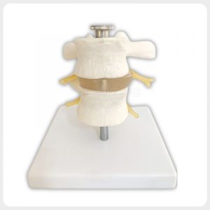
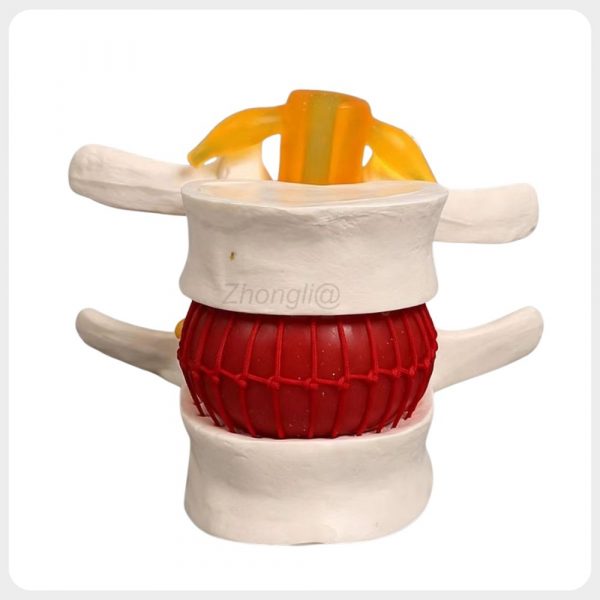
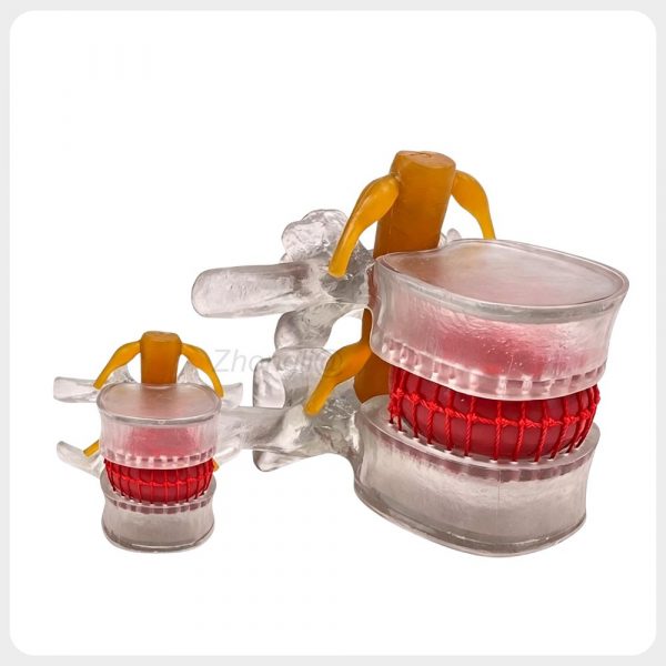
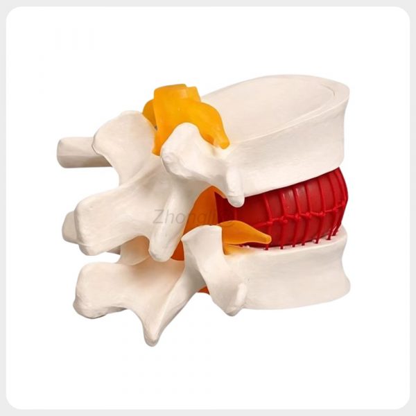
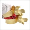
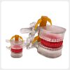
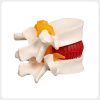
This compact lumbar disc herniation model clearly demonstrates the relationship between the vertebrae intervertebral disc protrusion and adjacent nerve roots. Three color options are available for medical teaching rehabilitation training and patient education.
1000 in stock
Product Description:
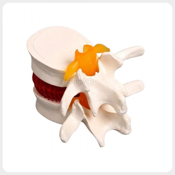
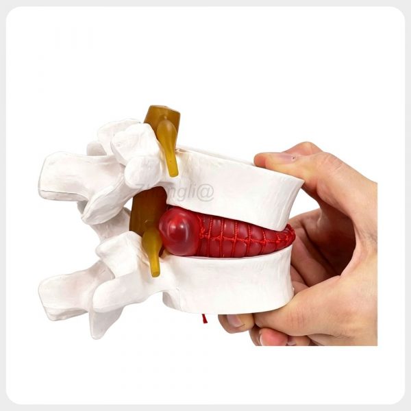
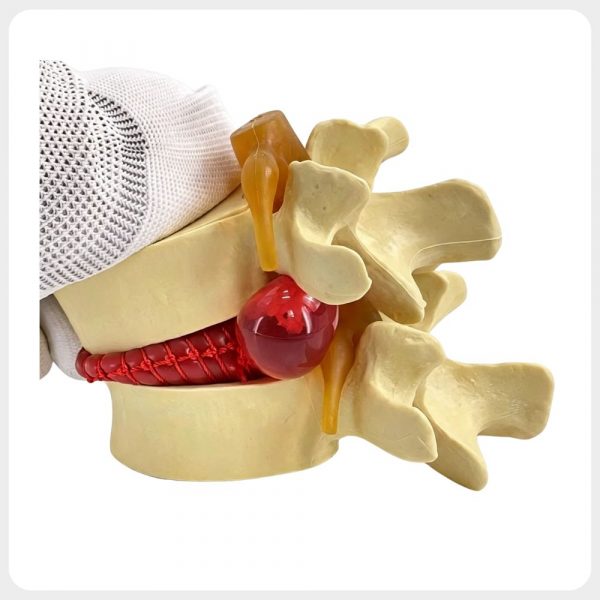
This anatomical lumbar disc herniation model presents two lumbar vertebrae a color coded intervertebral disc and adjacent nerve structures in a compact three dimensional format. The visible posterior disc protrusion helps students clinicians and patients understand how displaced disc tissue may approach or compress a nerve root.
Made of eco friendly PVC the model supports repeated demonstrations in medical schools orthopedic clinics rehabilitation centers and patient consultation environments. White transparent and natural bone color versions are available for different teaching and display requirements.
Key Features:
• Visible Disc Herniation: The enlarged posterior protrusion makes the relationship between displaced disc tissue and the adjacent nerve root easier to explain.
• Simulated Annulus Structure: The textured outer disc surface represents the fibrous annulus and helps explain how it normally contains the central nucleus.
• Color Coded Anatomy: Contrasting vertebra disc and nerve colors allow students and patients to identify important structures quickly.
• Compression Demonstration: The model can be handled during teaching to demonstrate how pressure relates to posterior disc protrusion and possible nerve contact.
• Compact Three Dimensional Design: The 11 x 10 x 7.6 cm model is suitable for clinic desks classroom demonstrations exhibitions and distributor sample cases.
Technical Specifications:
| Item | Specification |
|---|---|
| Product Name | Lumbar Disc Herniation Demonstration Model |
| Model | Lumbar Pathology Teaching Type |
| Brand | Zhongli |
| Material | Eco Friendly PVC |
| Scale | Anatomical Demonstration Scale |
| Product Size | Approximately 11 x 10 x 7.6 cm |
| Unit Package Size | Approximately 11.5 x 11 x 8 cm |
| Net Weight | Approximately 0.16 kg |
| Unit Gross Weight | Approximately 0.22 kg |
| Color Options | White Transparent and Natural Bone Color |
| Main Structures | Lumbar Vertebrae Intervertebral Disc Annulus Protrusion and Nerves |
| Application | Medical Teaching Patient Education and Rehabilitation Training |
| Carton Quantity | 64 Pieces |
| Outer Carton Size | Approximately 48 x 47 x 35 cm |
| Carton Gross Weight | Approximately 15.5 kg |
| Packaging | Individual Package and Export Carton |
Applications:
• Lumbar spine anatomy and pathology courses in medical nursing and healthcare schools
• Orthopedic spine pain management and neurology clinic patient education
• Rehabilitation physical therapy chiropractic and sports medicine training
• Medical equipment exhibitions distributor demonstrations and healthcare education programs
Packaging & Shipping

Packaging: The product is packed in a strong corrugated carton box to ensure safe transportation.
Inside the carton, the product is protected with foam or protective materials to prevent damage during shipping.
Standard export carton packaging is used to ensure the product arrives safely.

Global Shipping Options:
We support international express, air shipping, and sea shipping to ensure fast and reliable delivery worldwide.
Different shipping options are available depending on your order size and delivery time requirements
OEM & Customization
We support OEM and customized production based on customer requirements, including logo printing, color modification, packaging customization, and product structure adjustment.
Feel free to contact us for more details about private labeling and bulk order cooperation.
Get Our Latest Catalog
We also offer a full range of models in different sizes and structures. If you are interested, please feel free to contact us for our complete product catalog.
FAQ
Q1: What anatomical structures does this lumbar model demonstrate?
A1: It demonstrates two lumbar vertebrae an intervertebral disc the simulated annulus a posterior disc protrusion and adjacent nerve structures.
Q2: Can the model demonstrate the relationship between disc herniation and nerve compression?
A2: Yes. The posterior disc protrusion is positioned beside the colored nerve structure to help explain how displaced disc tissue may contact or compress a nerve root.
Q3: Can the model be used for compression demonstrations?
A3: Yes. The model can be handled and gently compressed during teaching to demonstrate the relationship between disc pressure posterior protrusion and possible nerve contact.
Q4: What colors are available?
A4: White transparent and natural bone color versions are available while the disc and nerve structures remain color coded for easy identification.
Q5: Is this model suitable for patient education?
A5: Yes. Its three dimensional color coded structure makes lumbar disc herniation easier to explain than verbal descriptions or flat images.
Q6: Is this product a diagnostic or treatment device?
A6: No. It is an educational model intended for anatomy teaching pathology demonstration rehabilitation training and patient communication.
| Product Type | Lumbar Disc Herniation Model |
|---|---|
| Product Size | 11 x 10 x 7.6 cm |
| Color Options | White Transparent and Natural Bone Color |
| Material | Eco Friendly PVC |
| Application | Medical Teaching and Patient Education |